Skip to content
Documented surgical cases

Clinical outcomes with digital planning context.

Explore selected periodontal, regenerative and implant surgery cases with diagnosis, planning logic, material context and realistic outcome notes. The goal is clarity, not exaggerated promises.

3D and clinical records

Cases are documented with photos, radiographs, CBCT or periodontal measurements when relevant.

Digital planning context

Implant, regeneration and soft tissue decisions are explained through the planning logic behind the surgery.

Materials and workflow

Each case clarifies guide use, implant system considerations, biomaterials or microsurgical technique when applicable.

Two-specialist review

Complex cases are positioned around specialist periodontal and oral surgical planning rather than generic dentistry.

Surgical documentation

Select a clinical focus.

Each case is educational. Treatment suitability, materials, guided surgery use and final outcome depend on diagnosis, anatomy, healing response and clinical examination.

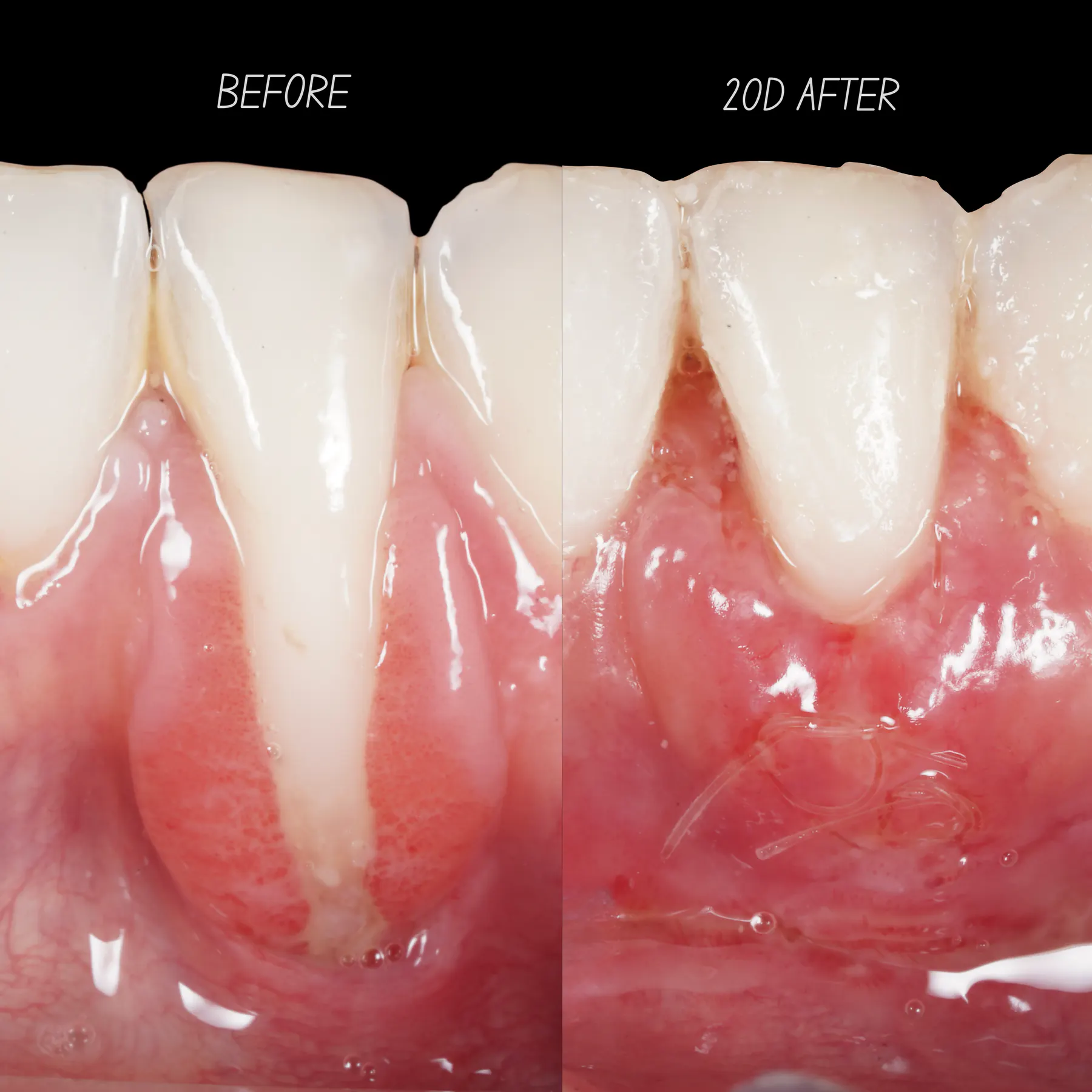
Gum recession before treatment and early healing at 20 days, as labelled in the clinical comparison

Recession Coverage

Severe recession root coverage

Connective tissue grafting followed by orthodontic care. See the initial assessment, the 20-day comparison and three-month photographs.

Photo protocolPeriodontal measurementsMicrosurgical planning

Assessment context

Photographic diagnosis
Periodontal measurements
View documented casePhotos: before, 20 days and 3 months. Invisalign-start records also shown.
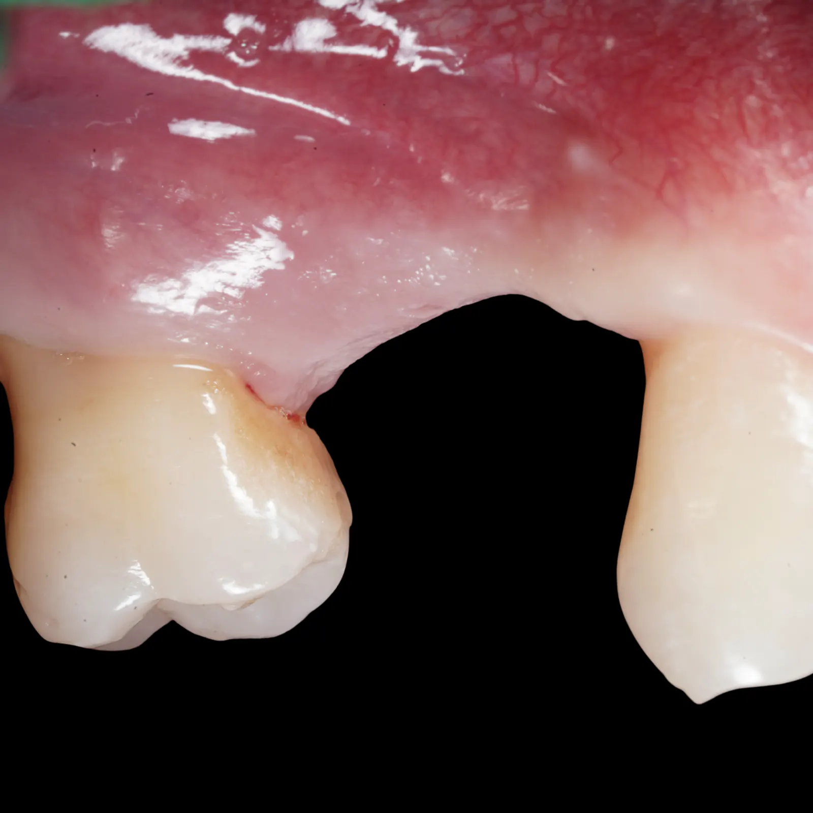
Clinical photograph of gum tissue across an upper posterior tooth space

Implant Surgery

Sinus lift and implant placement

A lateral sinus lift and implant placement in one operation, with healing assessment and the final restoration as separate stages of care.

CBCT planningImplant position planningRegenerative materials

Assessment context

CBCT sinus analysis
Prosthetically driven implant position
View documented caseSurface tissue photograph; healing assessed clinically and radiographically.
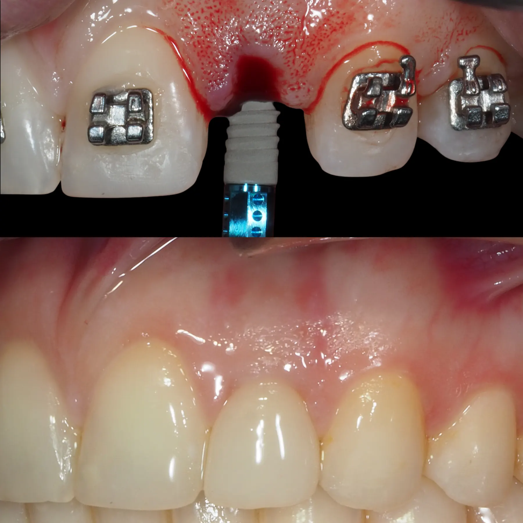
Anterior implant placement beside orthodontic brackets and a later restored tooth appearance

Multi-disciplinary

External root resorption and implant

Orthodontic space preparation followed by extraction, immediate implant placement and gum grafting, with a temporary tooth during healing before the final restoration.

Restorative-driven planAesthetic-zone implantSoft tissue design

Assessment context

Space and emergence planning
Aesthetic-zone implant position
View documented caseEarly healing at 2 weeks and final restoration delivery after approximately 3 months.
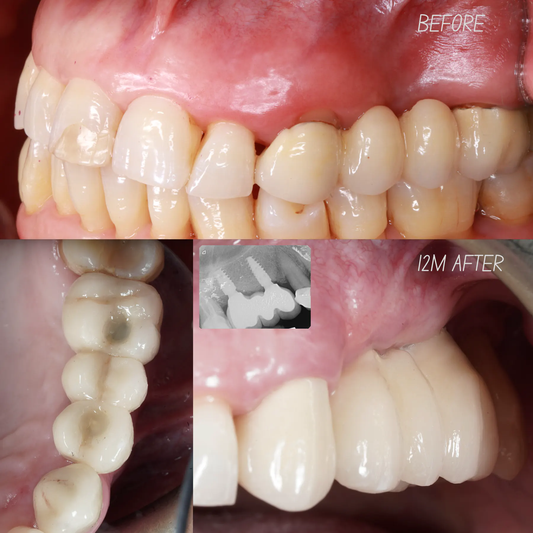
Clinical composite labelled before and 12 months after, with gum and tooth views and an inset implant radiograph

Bone Regeneration

Infection, regeneration and implant

Extraction and healing before implant placement with bone grafting, followed by restored views in a composite labelled “12M AFTER”.

Extraction and healingImplant with bone grafting12-month follow-up

Assessment context

Healing before reconstruction
Implant and graft together
View documented case12-month follow-up confirmed by the treating team, with clinical views and an implant radiograph.
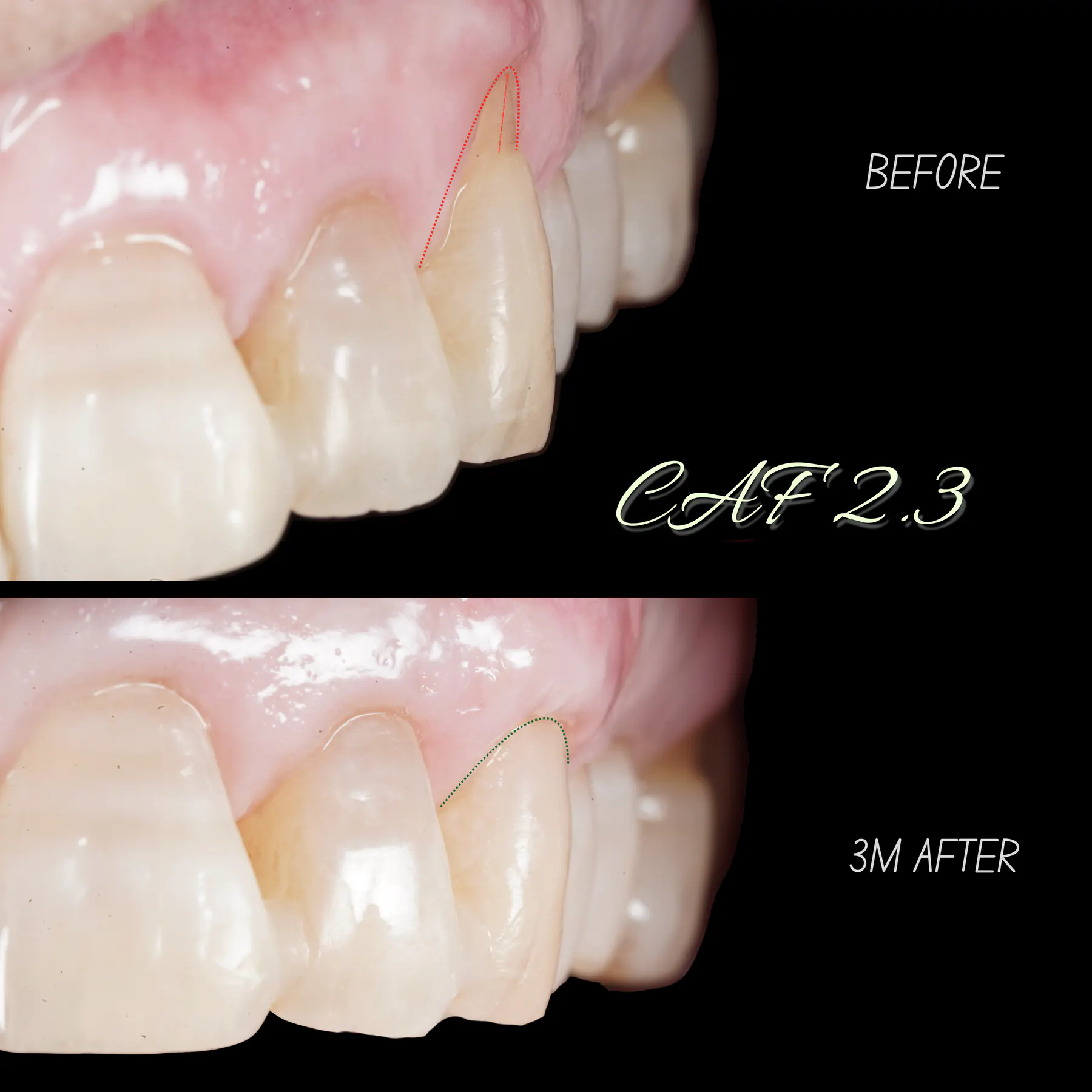
Upper left canine before corrective recession surgery and at three months, with original dotted outlines marking the gum contour

Recession Coverage

Corrective recession surgery

Corrective root coverage around an upper canine using a coronally advanced flap and connective tissue graft, with a three-month photographic comparison.

Corrective root coverageConnective tissue graft3-month photograph

Assessment context

Previous treatment context
Graft and flap together
View documented case3-month review confirmed by the treating team, matching the original photograph label.
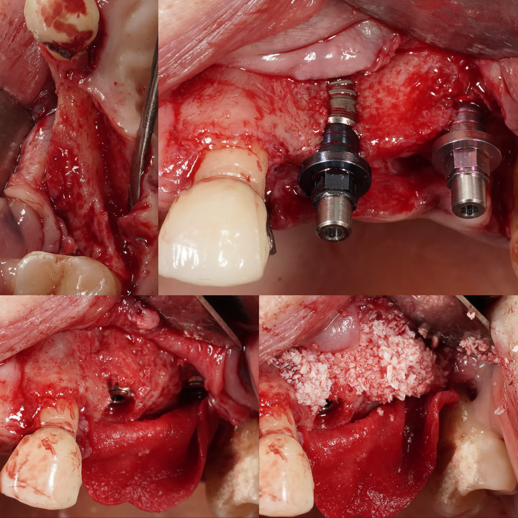
Four surgical views showing the exposed ridge, implants and placement of particulate bone graft material

Bone Regeneration

Bone and gum support after infection

Implant placement with horizontal bone grafting after extraction and healing, followed by a later roll flap to improve gum volume.

Horizontal bone graftingLater roll flapSurgical photographs

Assessment context

Outer bone wall assessment
Bone and gum in separate stages
View documented caseSurgical photographs shown; the team reports a radiographic review one year after crown rehabilitation.
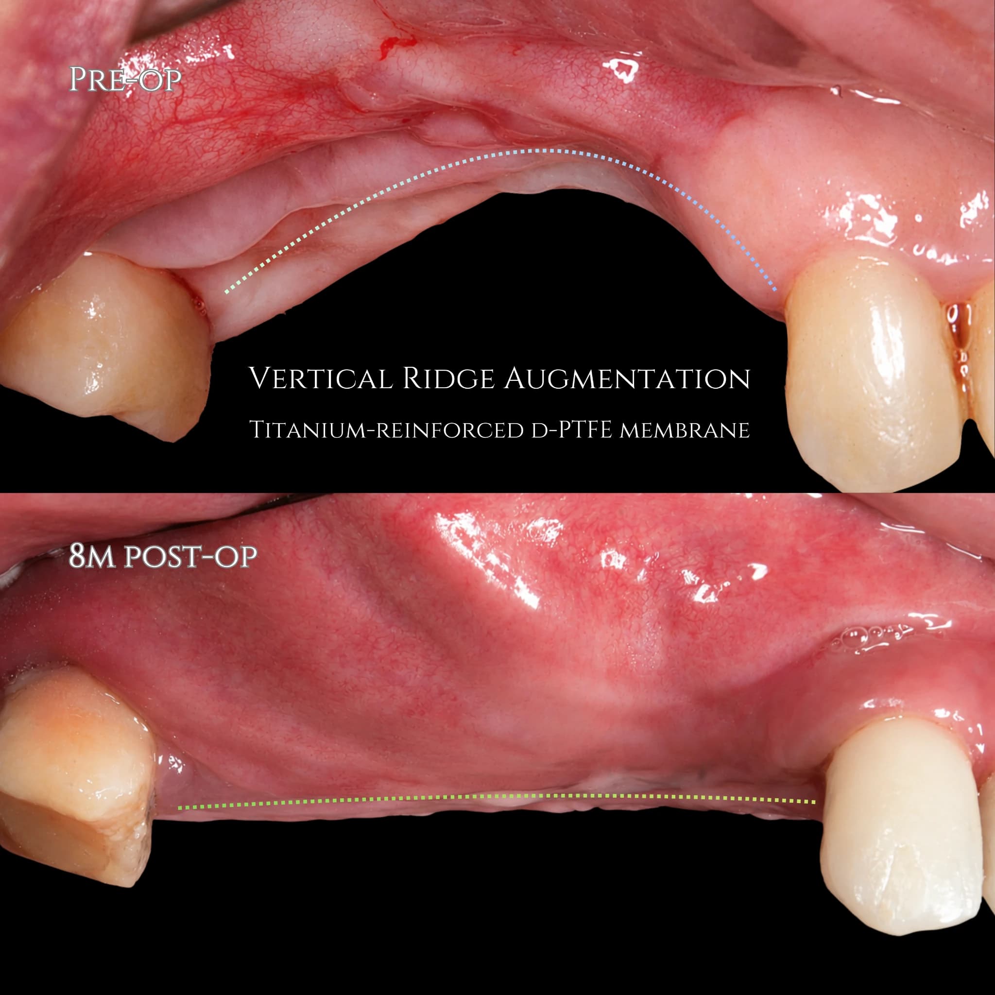
Upper jaw ridge before vertical augmentation and at eight months after surgery, with original dotted lines marking the visible gum contour

Bone Regeneration

Vertical bone regeneration

Staged reconstruction of lost ridge height after implant loss, with a reinforced membrane, an eight-month review and an illustrated guide to bone defects.

Defect morphology reviewStaged regeneration8-month review

Assessment context

Defect-driven diagnosis
Regenerative design
View documented case8-month surface comparison; subsequent implant placement confirmed, with surgical photos to be added.
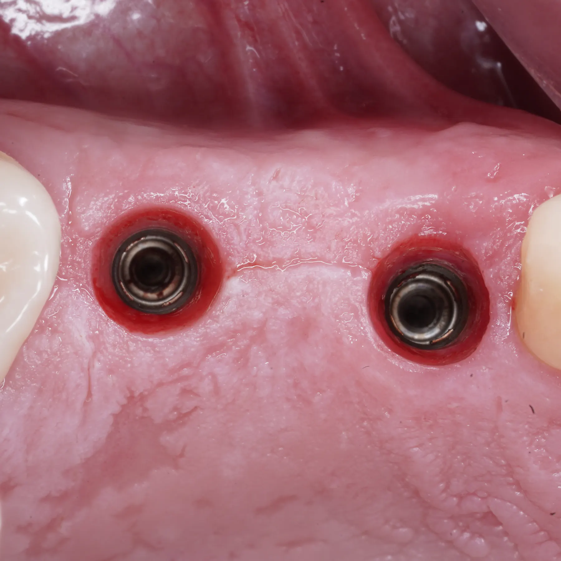
Close-up clinical view of gum tissue around two exposed implant connections, without crowns in place

Peri-implant

Immediate implants and surrounding gum

Extraction and immediate implant placement, with a close-up tissue photograph and an explanation of the surrounding gum.

Immediate placementTissue viewOngoing care

Assessment context

Confirmed treatment sequence
Reading the photograph
View documented caseSingle tissue photograph; review interval not specified.

Why this matters

Cases should show the decision-making, not only the final image.

Diagnosis

What problem was treated and why.

Planning

How records, digital design and surgical sequence guided the case.

Limitations

What factors influence predictability and healing.

Private assessment

Want to know whether your case is suitable?

Send your concern and available records before planning travel or surgery. The first step is understanding your case properly.

Private case review

Send your case before planning travel.

For international periodontal, regenerative and implant surgery enquiries.