Healthy tissues
No clinical inflammation. Bone support is stable.
Continue cleaning and regular reviews.
Compare the drawingsImplant health · Periodontal care in Barcelona
Peri-implantitis is inflammation with loss of the bone supporting an implant. We assess the cause and plan treatment to control it, aiming to keep the implant where possible.
Private care with Dr. Sergi Rivas Frau and/or Dr. Sahand Shirzadi Maryan, according to your treatment plan.
Visits take place at collaborating dental clinics in Barcelona. The location is confirmed when your appointment is arranged.
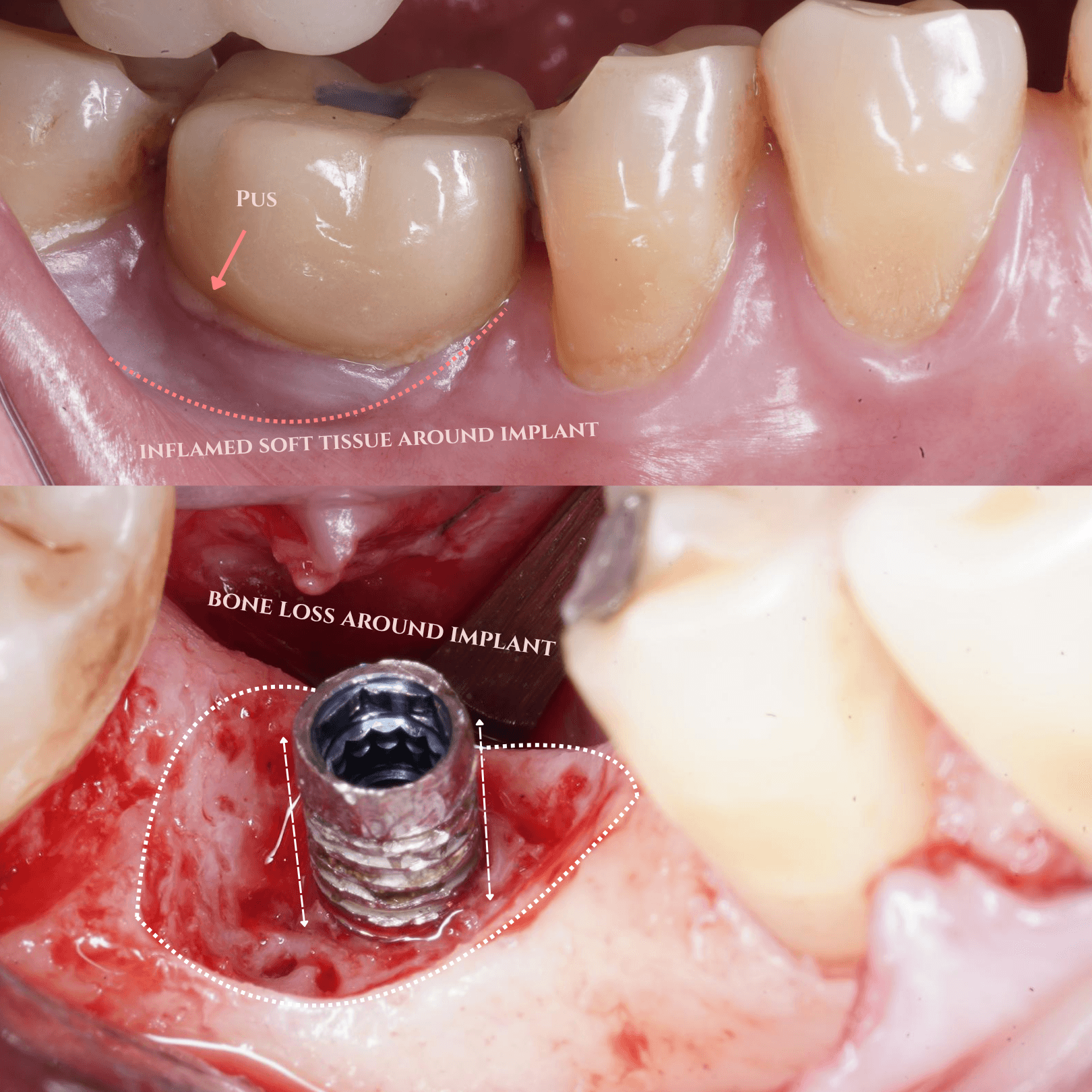
Clinical views of peri-implantitis
Upper view · before surgery: inflamed tissue around the implant.
Lower view · during surgery: after surgical cleaning (debridement), the implant surface and surrounding bone defect are visible.
The lower view shows an operative stage, not healing or a final result.
View full-size photographStart here · the whole journey
Your examination and response to initial care determine the next step. This map shows how the decisions fit together.
A reason to seek advice
Symptoms deserve an examination. They do not tell you on their own whether bone has been lost.
First · assessment
We check the gum, bone support, earlier X-rays and access for cleaning.
Different possible findings after examination
No clinical inflammation. Bone support is stable.
Continue cleaning and regular reviews.
Compare the drawingsInflamed gum without additional disease-related bone loss.
Professional cleaning, help with home care, then review.
Understand mucositisInflammation with progressive loss of the bone supporting the implant.
Initial cleaning and care for contributing factors, then review.
Follow the plan belowThese are alternative findings, not an inevitable sequence. The examination establishes which applies.
If peri-implantitis is diagnosed
Professional cleaning, home-care support and attention to relevant risk factors.
Check inflammation, measurements, cleaning access and bone stability where appropriate.
If disease is controlled
Continue cleaning and reviewsIf disease remains
Discuss whether surgery is suitableIf surgery is appropriate · options are chosen for your case and may be combined
Keeping the implant may sometimes be unsuitable. Removal and a replacement plan can then be discussed as an alternative.
Compare the tissues and bone support [1]
How to read the drawings
The same simplified bone-level implant is used throughout. In the intact-support example, the threaded body sits within bone and the gum meets the crown at its emergence.
Stable tissues
The tissues are free of clinical inflammation and the supporting bone is stable.
Inflammation + bone loss
Inflammation is present alongside progressive loss of the bone supporting the implant.
A different diagnosis
Mucositis affects the soft tissues. Peri-implantitis also involves disease-related bone loss. Examination and comparison with earlier records help distinguish them.[1]
These are different conditions, not inevitable stages. Health can also exist with reduced bone support; a low bone level alone does not establish active disease.
Peri-implantitis is associated with bacterial plaque around an implant; cleaning access, previous gum disease and individual health factors also matter.[1][4]
Bleeding, swelling, discharge or changes around an implant deserve assessment. Seek advice promptly if the implant feels loose or symptoms worsen. The extent of bone loss cannot be judged from symptoms alone.
From diagnosis to a treatment decision
The aim is to control disease and retain an implant where feasible. Initial care and the response to it guide further treatment.[3][4]
Step 01
We examine the tissues, measure around the implant and assess appropriate X-rays and earlier records. We also review your health, previous gum disease and the restoration.
Understand the implant, its support and the factors affecting care.
Step 02
Professional cleaning around the implant is combined with support for cleaning at home. Access beneath the crown or bridge and relevant risk factors are addressed.
Initial care prepares the tissues for reassessment and any further treatment.
Step 03
We review inflammation, tissue measurements, cleaning access and bone stability where appropriate. If control is adequate, continue cleaning and regular reviews. Persistent disease may need a surgical approach.
The response to initial care helps determine the next step.
If control is adequate
Continue cleaning and reviewsIf disease persists
Discuss surgical optionsBone-defect patterns · Monje and colleagues
Bone loss can have different configurations. These anatomical patterns describe the defect; they do not prescribe treatment or represent a sequence of stages.[2]
Class I · infra-osseous
A defect extends into the remaining bone. The number and shape of the bone walls vary.
Class II · horizontal
Bone support is reduced across the crest, leaving more of the implant above the remaining bone.
Class III · combined
A lower crest and a deeper defect are present together. Different approaches may be combined.
We also assess remaining bone walls, open-sided defects and how the implant sits within the bone. Containment may matter when considering reconstruction, alongside inflammation control, tissue conditions and the restoration.
Different options for different needs
Surgery can provide access to affected areas. The approach depends on the defect, cleaning access and treatment goals; techniques may be combined.[3][4][5]
Option A · selected after assessment
The gum is gently opened to reach affected areas, remove inflamed tissue and clean the implant surface.
When considered: When deeper access is needed to address persistent disease.
Discuss the limits: The gum position can change after surgery; some implant surface may become more visible.
Option B · selected after assessment
Selected gum tissue and bone may be reshaped or removed to improve cleaning access and reduce difficult-to-maintain pockets.
When considered: When the anatomy and restoration make this approach appropriate.
Discuss the limits: Recession, exposed metal and changes in appearance need discussion, especially in a visible area.
Option C · selected after assessment
Graft material, sometimes with a membrane, may be used to support reconstruction of a suitable defect after cleaning.
When considered: Defect depth, remaining bone walls and tissue conditions help guide suitability.
Discuss the limits: A graft does not guarantee new bone attachment or implant rescue. Further care may still be needed.
Option D · selected after assessment
If keeping the implant is no longer a reasonable option, removal and a separate replacement plan may be discussed.
When considered: The decision considers support, stability, persistent disease and the overall restoration.
Discuss the limits: There is no single bone-loss percentage that decides this for every patient.
Cleaning methods and materials are selected for the case. No single device guarantees disease resolution. Radiographic bone fill does not by itself prove new bone attachment to the implant or lasting disease control.[3][4]
Long-term care is part of treatment
Peri-implantitis can recur. Clear cleaning advice and regular professional reviews help identify changes and guide further care. Follow-up is normally with our periodontist team.
Ask how tissue measurements and bone levels will be monitored, what changes to report, and how reviews will be arranged if you live abroad.
Patient understanding and realistic expectations matter. [6]
Your questions
Ask for the proposed sequence, alternatives, costs and follow-up in your written treatment plan.
Bleeding needs assessment. Gum inflammation alone can be mucositis; peri-implantitis also involves disease-related bone loss. Examination and records help distinguish the conditions.[1]
No. The response to initial care, the defect, cleaning access and other findings determine whether surgery or reconstruction is appropriate.[2][3]
Our aim is disease control and implant preservation where feasible. Retaining an implant is not possible or appropriate in every case.[4]
Yes. Improvement needs ongoing review. Home care, professional maintenance and checking for recurrent inflammation are part of the long-term plan.[6]
Treatment is planned around the diagnosis and contributing factors. Antibiotics are not a routine substitute for cleaning and appropriate further care; no single device guarantees disease resolution.[3][4]
Yes. Contact us with a brief description and any available records. Suitability and the sequence of visits need assessment before you arrange treatment or travel.
The evidence behind this guide
Patient explanations are informed by consensus guidance and peer-reviewed research, including Monje’s work on defects, combined treatment and patient understanding. Research findings do not predict an individual outcome.
[1] Berglundh et al. · 2018
Peri-implant diseases and conditions: World Workshop consensusJournal of Clinical Periodontology
[2] Monje et al. · 2019
Morphology and severity of peri-implantitis bone defectsClinical Implant Dentistry and Related Research
[3] Herrera et al. · 2023
Prevention and treatment of peri-implant diseases: EFP S3 guidelineJournal of Clinical Periodontology
[4] Wang et al. · 2025
AO/AAP consensus on prevention and management of peri-implant diseases and conditionsJournal of Periodontology
[5] Monje et al. · 2026
Extent of implantoplasty in combined surgical therapy: a quasi-randomized clinical trialClinical Implant Dentistry and Related Research
[6] Monje et al. · 2026
Understanding peri-implantitis occurrence and recurrence following treatment: a patient-centered studyJournal of Periodontology
Patients in Barcelona and abroad
Tell us briefly what you have noticed, what you have been advised and whether previous records are available. An assessment establishes the diagnosis and a plan for your case.
An enquiry does not confirm an appointment or treatment suitability. Contact us before booking travel.
Private case review
Send your case before planning travel.
For international periodontal, regenerative and implant surgery enquiries.