Implant tissue correction
Peri-Implant Soft Tissue Reconstruction
Soft tissue correction around implants to improve tissue thickness, keratinized mucosa, aesthetics and hygiene access in compromised implant sites.
Implant tissue correction
Surgery is designed before the surgical appointment begins.
Designed for
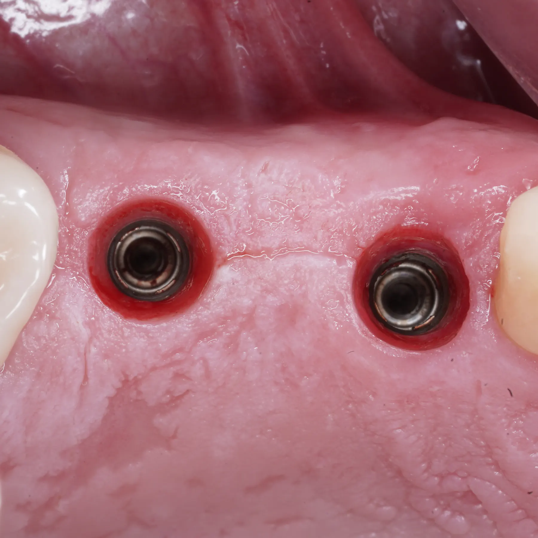
Thin mucosa around implants
Implant recession or visible grey shadow
Insufficient keratinized tissue around implants
Soft tissue problems affecting hygiene or aesthetics
Planning records
Evaluation of implant position and prosthetic contour
Assessment of inflammation, peri-implant pocketing and bone levels
Soft tissue phenotype and keratinized tissue measurement
Coordination with restorative correction when needed
Clinical sequence
How this treatment is approached.
Diagnosis
Implant sites with thin tissue, recession, visible metal or difficult hygiene access may need soft tissue reconstruction to improve stability and comfort.
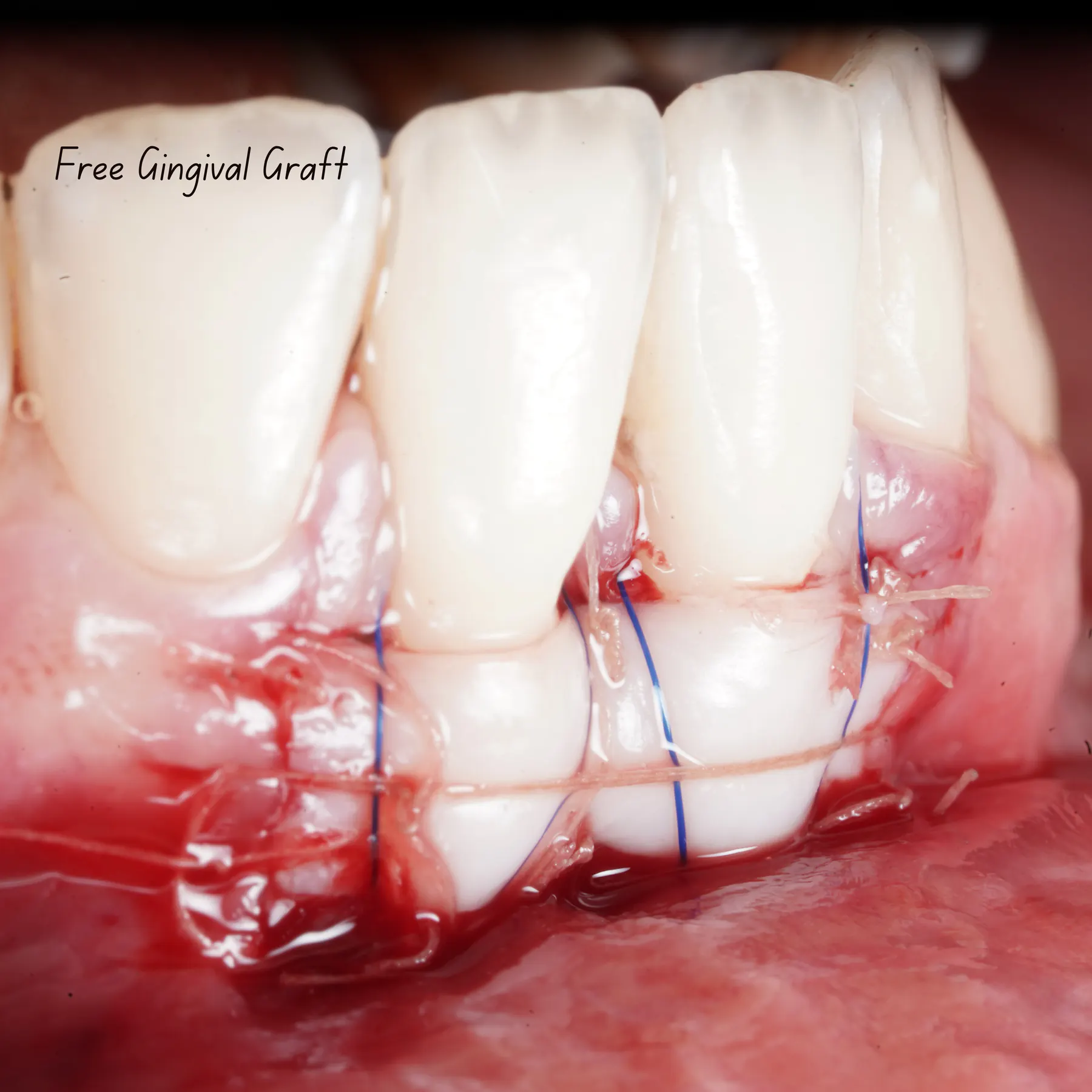
Surgical approach
Connective tissue grafting or keratinized tissue augmentation Flap design adapted to implant position and soft tissue needs Prosthetic contour modification if tissue health requires it Maintenance planning for long-term peri-implant health
Digital advantage
Photographic documentation clarifies soft tissue changes over time Digital planning helps evaluate implant/prosthetic factors behind tissue problems Remote review can identify records needed before travelling
Technology and materials
Microsurgical soft tissue instruments Autogenous grafting and selected biomaterials when indicated Digital photo and record-based follow-up planning
Digital workflow value
Photographic documentation clarifies soft tissue changes over time
Digital planning helps evaluate implant/prosthetic factors behind tissue problems
Remote review can identify records needed before travelling
Materials and technology
Microsurgical soft tissue instruments
Autogenous grafting and selected biomaterials when indicated
Digital photo and record-based follow-up planning
Important limitations
Precision requires honesty.
Premium surgical planning improves clarity, but treatment suitability and outcome depend on diagnosis, anatomy, tissue quality, healing response and clinical execution.
Limitations to respect
If the implant is severely malpositioned, tissue grafting alone may not solve the problem
Inflammation and plaque control must be addressed before or alongside surgery
Final aesthetic correction depends on bone, implant position and prosthetic design
Patient pathway
Send close-up implant photos and any X-rays
Clarify whether there is bleeding, pain, recession or aesthetic concern
Plan soft tissue correction alone or as part of broader implant retreatment
Clinical information and outcome notice
The information on this website is educational and does not replace an individual diagnosis, clinical examination, radiographic assessment, periodontal charting, CBCT review, or personalised medical advice. Treatment suitability and outcomes vary according to anatomy, health status, diagnosis, healing response, compliance, and other clinical factors.
Related treatment planning
Complex cases often combine several disciplines.
Tissue architecture
Soft tissue grafting
Soft tissue grafting to improve tissue thickness, keratinized tissue, peri-implant stability and aesthetic tissue architecture around teeth or implants.
Explore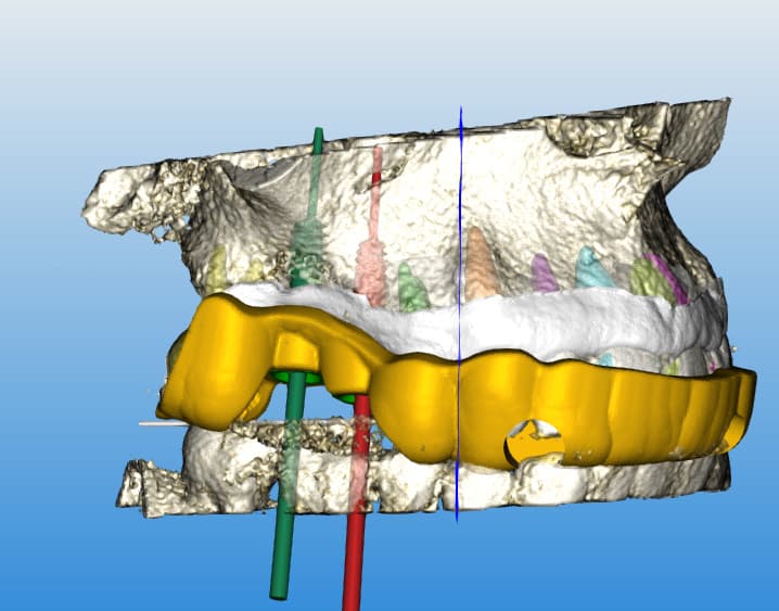
Digital implant planning
Guided implants
A digitally planned implant workflow using clinical records, CBCT information, intraoral scans and guide design to support accurate implant positioning when guided surgery is clinically indicated.
ExplorePrivate case review
Send your records before planning treatment.
The first step is to understand your anatomy, diagnosis, expectations and travel context before defining a surgical sequence.