Observation, clinical case mentorship and focused workshops for qualified dentists in Barcelona. Prepare through scientific articles, theory and complex-case planning.
Learn with Dr. Sahand Shirzadi Maryan and Dr. Sergi Rivas Frau. Connect theoretical foundations with periodontal, regenerative, implant and restorative decisions.
Arranged individually. We confirm eligibility, content, teaching language, dates, duration and fees before you make arrangements. An enquiry does not reserve a place.
Both clinicians completed master’s training in periodontology and implant dentistry at the University of Barcelona.
These are the clinicians’ qualifications; they do not indicate university accreditation of Academy programmes.
Find your starting point
Different ways to learn. A clear role in each.
You may want to observe a procedure, discuss a treatment plan or focus on a specific technique. Your qualifications, experience and objectives help us decide which format may fit. Clinical participation is a separate arrangement.
Advanced Surgical Observation
Qualified dentists and postgraduate clinicians seeking to observe advanced surgery.
Learning focus
Prepare through articles and theory, review the complex treatment plan before surgery, then connect the observed procedure with the clinical decisions.
For now, surgical observation, case mentorship and focused workshops are arranged individually with our team. We confirm eligibility, content, teaching language, dates, duration, location and fees before you make arrangements. An enquiry does not reserve a place.
Live observation depends on the case, schedule and patient consent. Workshop exercises or observation are included only when explicitly agreed.
Looking for treatment as a patient? Our planned supervised-care route has its own explanation of who provides treatment and how supervision works.
Read the evidence. Plan the case. Understand the surgery.
Learning starts before the surgical setting. We use journal articles and theoretical discussion to build the foundations, then show how those principles inform a complex treatment plan. The focus is the whole patient and intended restoration, not an isolated technique.
01
Build the foundations
What supports this approach?
Read selected journal articles and revisit diagnosis, anatomy, case selection and risk assessment. Discuss the strength and limits of the evidence behind established and evolving techniques.
02
Plan the complex case
How do the decisions fit together?
Before surgery, review the records, treatment alternatives, digital planning when relevant and the intended restoration. Understand the reasons for the sequence, staging and contingency decisions.
03
Connect plan and practice
How is the reasoning applied?
In observation, follow the agreed surgical case from an observer position. In mentorship or workshops, work through case decisions and focused learning activities. Participation follows the chosen format.
04
Debrief and reflect
What will you take back to practice?
Relate what you have seen or discussed to the original plan and articles. Explore limitations, healing assessment and further reading, and identify questions for continued learning.
Explore our documented cases and educational illustrations for an introduction to this way of thinking. They explain the principles; they are not surgical instructions or a complete training course.
Learn how to connect clinical records, the intended restoration and a surgical plan. Explore the decisions behind implant positioning and guide design with Dr. Sahand Shirzadi Maryan and Dr. Sergi Rivas Frau.
Our teaching also explores digital planning for sinus access and horizontal and vertical bone reconstruction, with the approach selected around the clinical problem.
Inside a planning case
1 min 24 sec · No narration
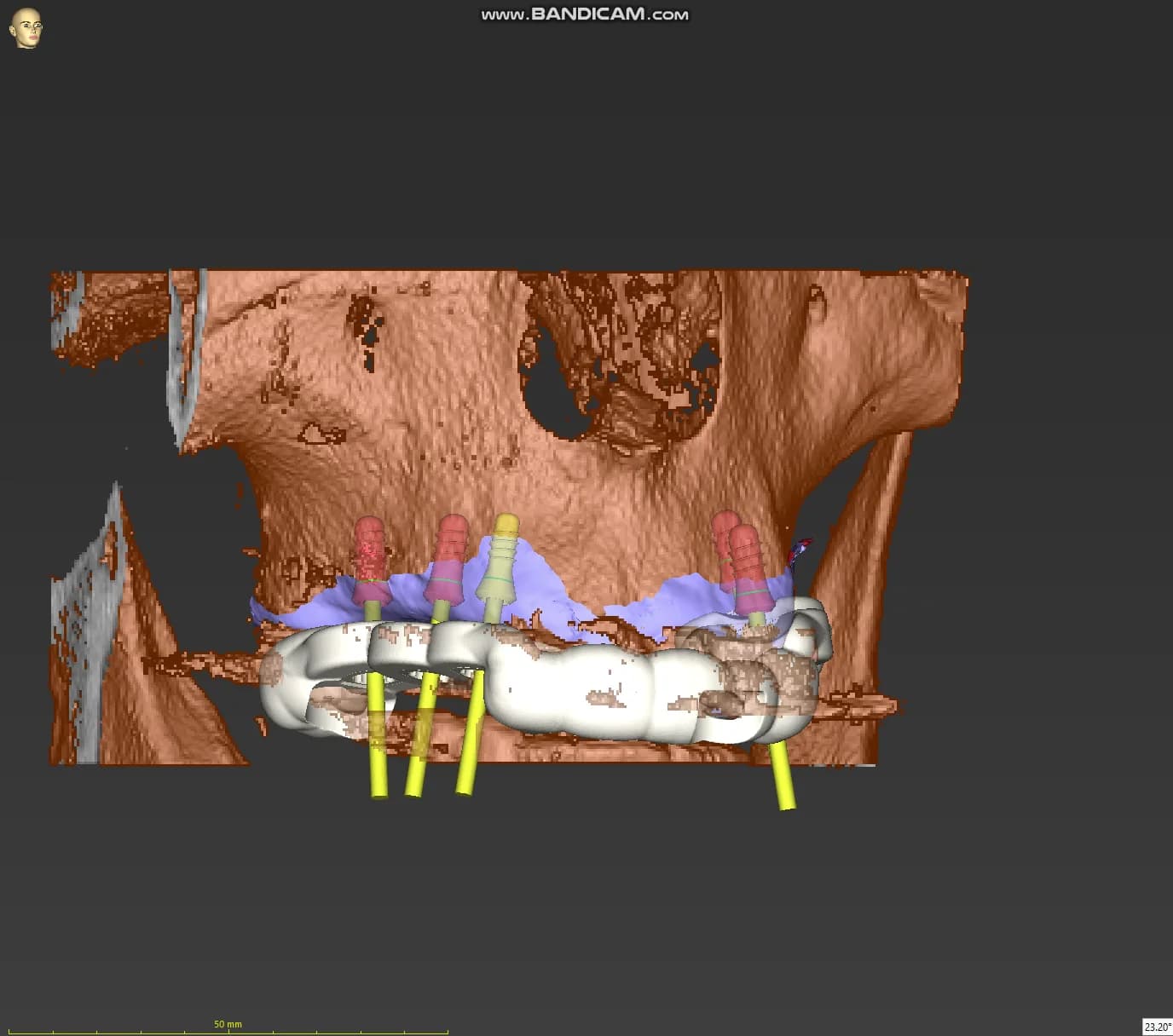
Two upper-jaw implant bridges: three implants + two implants.
The rotating coDiagnostiX view shows the plan for two separate upper-jaw bridges: one supported by three implants and the other by two. This is a partial-arch restoration, not a full-arch bridge. Surface scans, bone anatomy and restorative and guide components help explain the planning relationships.
A digital planning demonstration. The implant number, guide design and treatment stages are specific to the individual case.
What to look for
Follow the reasoning behind the plan.
01
The records together
How do the surface scan, bone anatomy and proposed teeth relate to each other?
02
The intended restoration
How does the restorative goal inform the position and distribution of the planned implants?
03
The guide and its purpose
Which parts of the agreed plan should the guide help transfer, and what still needs clinical verification?
Prepare. Plan. Discuss.
Selected articles and theoretical preparation give the case its context. We discuss indications, alternatives, limitations and how the planning decisions apply to your own clinical practice.
The learning pathway
Connect each step with the next.
01
Read the records
Connect the examination, photographs, surface scans and appropriate three-dimensional imaging.
02
Define the restoration
Start with the intended teeth, function and cleaning access, then consider their supporting tissues.
03
Build the digital plan
Discuss implant positions, the available anatomy and whether regeneration or separate stages are needed.
04
Design and verify the guide
Understand its purpose, support and fit, and how the design relates to the agreed surgical plan.
05
Connect the plan with care
Relate planning decisions to clinical checks, surgical judgement and the subsequent review.
A clinical teaching case · Orthodontic collaboration
Guided corticotomy. One coordinated orthodontic plan.
The orthodontic department referred this patient for a surgical stage intended to assist expansion of the upper dental arch before planned orthognathic surgery. The expansion appliance had been prepared with the orthodontic team before surgery.
The surgical plan combined bilateral posterior corticotomies with bone grafting and a membrane, aiming to increase alveolar bone volume in the same regions. These records follow the separate guides into clinical use and an early two-week review, while expansion treatment continued.
Surgery
Dr. Sahand Shirzadi Maryan
Guide design & printing
Dr. Behnam Taghavi
01Check the model and guides
02Transfer the planned markings
03Remove the guide and follow the markings
04Review alongside orthodontics
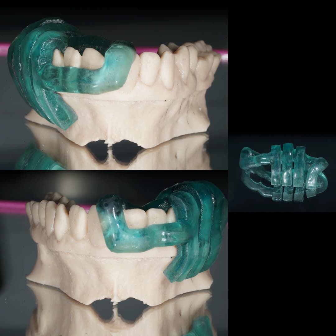
01 · The printed model and two separate guides
The two large photographs show the guides on the printed maxillary model, one for each side. The smaller view shows one guide by itself. This is a model check, before placement in the mouth.
The guides, designed and printed by Dr. Behnam Taghavi, relate the intended markings to the patient’s teeth. The model lets the team inspect their design and seating before the clinical stage. Fit is checked again in the mouth.
A marking guide, followed by direct surgical control
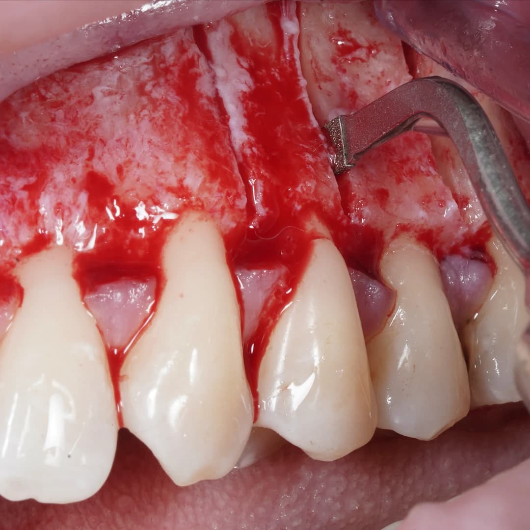
The guide transfers the planned locations. It is then removed before the piezoelectric instrument follows those markings under direct vision, with depth controlled separately.
For discussion: how do guide support, seating, root positions and the planned movement influence the design and its clinical checks?
View surgical photographs · stages 02–03Contains exposed gum and bone. Open to follow the guide placement and piezoelectric stage.
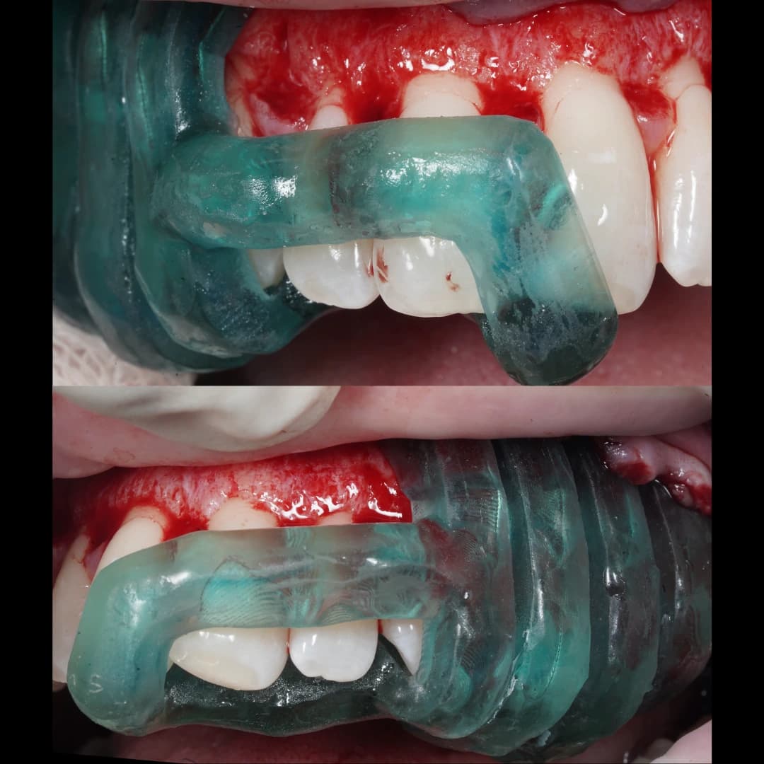
02 · Each guide seated in the mouth
Upper photograph: the guide on the patient’s right side. Lower photograph: the guide on the patient’s left side.
The guides are used in turn to transfer the planned markings. These views document seating, rather than the depth of the subsequent cuts.
After marking, the guide is removed. The piezoelectric instrument follows the transferred lines in the exposed cortical bone, with depth controlled clinically according to the site and plan.
Bone grafting with a membrane was also performed in the treated posterior regions on both sides, aiming to increase bone volume. Graft placement is not shown in this photograph.
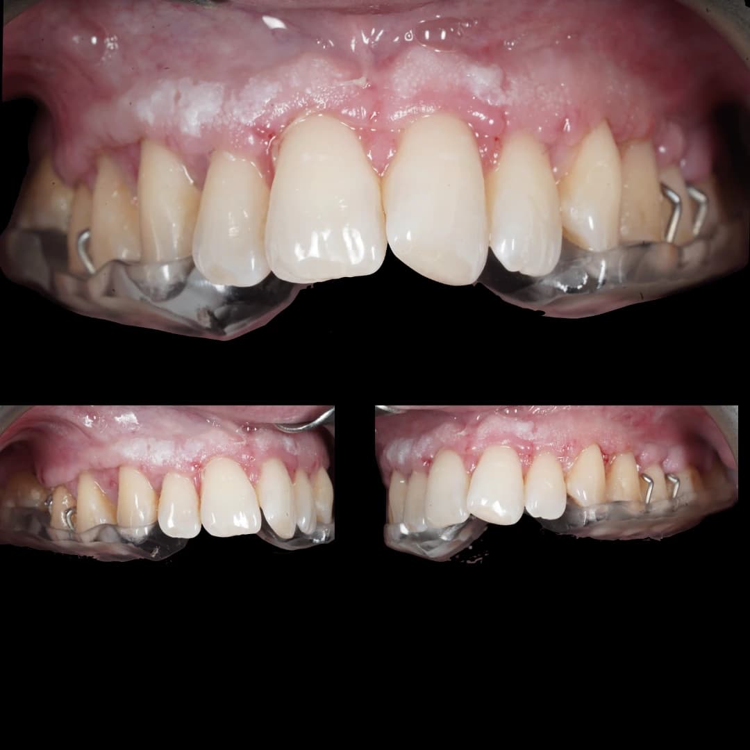
At the two-week review, the patient was continuing with the expansion appliance prepared before surgery by the orthodontic department. The composite photograph shows front and side views at that visit.
The review records an early postoperative stage. It does not measure bone regeneration, the amount of skeletal expansion, long-term stability or the eventual orthognathic result.
The shared treatment plan remains central.
Appliance use, activation and later orthodontic or jaw-surgery stages are directed by the treating team. This case is not a general indication for corticotomy in every patient who needs expansion.
Two-week review · expansion appliance in use
An early follow-up record while orthodontic treatment continues. It is not a final treatment comparison.
Journal of Indian Society of Periodontology. 2018;22:328–333.
A clinical comparison using a different indication. The reported cortical grooves were approximately 1.5–2 mm deep; this is not a depth recommendation for the case shown here.
Journal of Oral Biology and Craniofacial Research. 2021;11:63–65.
A technique report describing approximately 3 mm corticotomies with grafting through a tunnel approach. Its access and technique differ from the open surgical views in this case.
A printed-model study found greater accuracy for several measurements with guides, but not depth. It does not establish the accuracy or safety of this patient’s guides or the marking workflow shown here.
This documented sequence is for patient understanding and professional discussion. It does not replace case-specific diagnosis, surgical training or clinical supervision.
Bring your learning goals into the discussion.
Tell us your clinical experience and where you want to use digital planning. We can discuss an individually arranged workshop or mentorship, with content and practical activities agreed in advance.
For qualified dentists. Dates, teaching language, content and fees are confirmed individually. Observation and practical exercises are included only when agreed; supervised patient treatment remains a separate programme in preparation.
Programme formats
Choose the educational format that matches your clinical level.
Tell us about your experience and learning goals. We will confirm available formats, eligibility, dates, duration and fees before you make arrangements. Supervised patient treatment is still in preparation.
Further training. With supervision at the patient’s side.
We are preparing a programme for qualified dentists undertaking further clinical training with OsteoPerio. The planned format involves selected patient treatment with Dr. Sahand Shirzadi Maryan and Dr. Sergi Rivas Frau both present during surgery.
This is a future clinical participation format. Observation and case mentorship do not themselves include performing treatment. The supervised programme is intended for already-qualified dentists, rather than undergraduate dental students.
The planned roles
Who would do what?
The dentist in training
An already-qualified dentist would undertake selected treatment within the agreed scope of training and supervision.
Both supervising clinicians
Sahand and Sergi would both be present during surgery to supervise the clinical work.
Continuity after surgery
Follow-up would normally be provided by our periodontist team. The review visits and responsibilities would be agreed for each patient’s treatment.
This describes the planned model. It does not confirm eligibility, a training place or access to a particular patient or procedure.
Participation starts with a suitable match.
A dentist’s qualification alone does not determine which procedures they may undertake. Their experience, the patient’s needs, case complexity and the proposed supervision all need to be considered together.
01
Professional background
Qualifications, current role, relevant surgical experience and learning objectives would be reviewed before any participation is agreed.
02
Patient and case suitability
Case selection, the treatment plan, the proposed operator and patient consent would need to be agreed before treatment. Educational goals must fit the patient’s care.
03
Defined responsibilities
The permitted clinical role, supervision, clinical setting and follow-up arrangements would need to be confirmed for the proposed training.
What remains to be confirmed?
The programme’s start date, duration, teaching language, fees and inclusions, detailed entry requirements and clinical arrangements have not yet been finalised. No specific case numbers or procedures are promised.
Any participation documentation would also need to be confirmed. The clinicians’ university qualifications do not mean that this programme awards a university degree or specialist qualification.
Ask about the planned programme.
Dentists can send a professional enquiry with their training background, experience and learning interests. An enquiry is not a booking, a reservation or confirmation of clinical participation. Please wait for confirmed arrangements before planning travel.
Your selected format is preselected in the education enquiry. You can change it there; an enquiry does not reserve a place.
Looking for treatment as a patient?
The planned reduced-fee patient route has its own explanation of treatment, supervision and follow-up. It is also in preparation and unavailable to book.
Every learning format respects the patient, the clinical setting and each participant’s agreed role.
Patient consent and clinical suitability guide observation and case use.
Case discussions use appropriately anonymised records.
Clinical priorities and the treating team’s directions come before educational activities.
Explore learning topics
Topics depend on the agreed format and your learning goals.
Periodontal diagnosis, phenotype assessment and surgical case selection
Clinical photography, CBCT review and digital documentation workflows
Soft tissue grafting around teeth and implants
Bone regeneration, ridge preservation and staged implant planning
Guided implant surgery, guide splints and prosthetically driven planning
Sinus lift and posterior maxilla treatment sequencing
Complication prevention, patient communication and follow-up planning
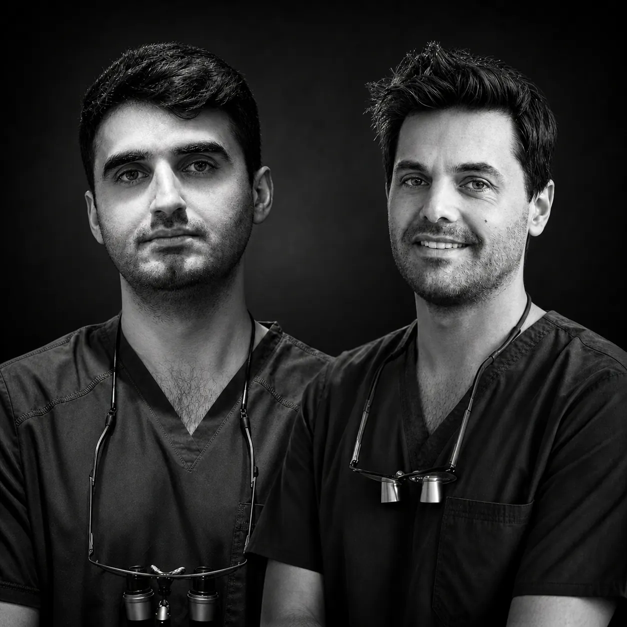
Dr. Sahand Shirzadi Maryan Dr. Sergi Rivas Frau
Practical questions
Before you enquire.
Who can enquire?
Qualified dentists, including those in postgraduate training, can enquire. Share your experience and learning goals so we can assess which format may suit you.
How are Academy programmes arranged?
For now, all programmes are arranged individually. Tell us your professional background, learning goals and preferred dates; we will discuss a suitable format and confirm the content, duration, teaching language, location and fee before you decide.
Can I perform surgery?
The supervised clinical programme is in preparation and not bookable. It is planned for qualified dentists, with both Sahand and Sergi present during surgery. Observation, mentorship and workshops do not include treating patients.
Is live surgery observation included?
Only if agreed for your format and permitted by the case, schedule and patient consent. Observation is not included in every programme.
Can I attend from abroad?
International dentists can enquire. Confirm eligibility, teaching language, dates and clinical location with the team before booking travel.
How long is a programme, and what does it cost?
Duration and fees are confirmed for the agreed format, content and arrangements. Tell us your goals and preferred timing. Before you commit, clarify what is included: preparation, teaching time, materials and any follow-up.
Which language will the teaching be in?
Confirm the teaching language with the team before arranging your programme or travel. The website language and written-enquiry language do not confirm the language used for teaching.
Are certificates or qualifications awarded?
Ask what participation documentation is included. The clinicians’ university qualifications do not imply that Academy programmes are university-accredited or award a specialist qualification.
Your next step
Tell us what you want to learn.
Share your professional background, preferred format and learning goals. For the first enquiry, please leave out patient-identifying details and clinical files.
01
Share your goals
Tell us your experience, interests, preferred timing and teaching language.
02
Discuss the fit
We review suitability and which formats may be available.
03
Confirm the details
Agree the content, dates, location, fees and participation requirements before making arrangements.